
3D imaging technology changes surgical plan and life for OSF patient
Key Takeaways:
- 3D imaging which allowed for multi-dimensional viewing of a tumor changed the surgical plan for an OSF patient with a rare lung cancer.
- The technology allowed the surgeon to avoid removing three ribs, leading to an easier, safer recovery from tumor removal and with minimal pain.
- The patient Jake Wilson now promotes hope and an annual lung CT screening, crediting early detection and his faith.
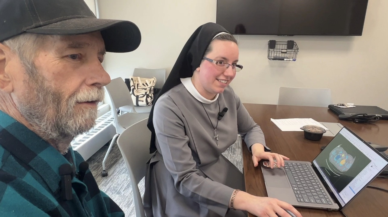
A new generation of 3D Virtual Reality (VR) imaging technology at OSF HealthCare is giving surgeons a clearer view of cancer than ever before – and in some cases, completely changing surgical plans. For OSF HealthCare thoracic surgeon Richard Anderson, MD, that level of insight recently made the difference between removing part of a patient’s chest wall and preserving it entirely.
Dr. Anderson, chair and Norman Estes Professor of Surgery at University of Illinois College of Medicine Peoria, says the leap from standard imaging to 3D modeling is transformative for surgeons.
A better view
“So the real difference between a 2D and 3D is, just imagine looking at something on a piece of paper with two different pictures, looking at it from side to side, as opposed to having a model in your hand and being able to take that model and move it all around in all different directions, upside down, inside out, even open up things within the model and look at it. It’s really the difference between 2D and 3D.” Dr. Anderson adds, “It’s really quite impressive.”
The technology allows his team to isolate tumors, peel away layers of anatomy, and examine the intricate relationships among bones, vessels, and muscle before they ever enter the operating room. That clarity is especially crucial in complex thoracic oncology, where anatomy is delicate, variable and easily injured.
For Dr. Anderson and others who use 3D imaging, it increases the surgeon’s confidence.
“In the more complex oncology cases, it can make a big difference. It can help the surgeon really identify what structures they have to worry about during the operation, what structures they might have to sacrifice, before they actually do the operation and gives them a little bit more level of confidence going into the operation than probably they would have before.”
A tumor in a rare location
That level of detail proved critical in a recent complex thoracic oncology case. Thoracic surgery presents unique challenges because of the delicate and complex anatomy of the chest.
“The arteries aren't made the same, and so they're easier to tear. They're much more delicate. Lung tissue is sometimes like tissue paper, too. It’s easy to injure and tear. You have got to be very careful when you do the dissections in thoracic surgery. That's what makes it so challenging.”
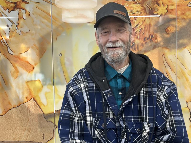
Dr. Anderson treated Jake Wilson, a 69-year-old retired farmer and former prison guard from Central Illinois. Wilson’s case was challenging. He was diagnosed with a rare form of lung cancer and in August of 2025 began a series of chemotherapy and radiation to shrink the aggressively growing tumor.
Still, as surgery approached, Dr. Anderson was challenged with removing a nearly six‑centimeter tumor (about the size of a small egg) situated high in the chest, closely hugging the first three ribs. Standard 2D scans suggested the cancer had invaded the bone – a scenario that would typically require removing three ribs to ensure the cancerous tumor was completely removed.
 Seeing is believing
But when Wilson became the first adult patient with lung cancer to have his case analyzed using advanced 3D modeling and virtual reality, the picture changed dramatically. The technology revealed that his ribs were not invaded at all. The tumor, though large and aggressive, was confined to muscle.
Sister M. Pieta Keller, Connor Davey, and Reid Jockisch, innovation engineers at Jump Simulation worked to segment the tumor for 3D viewing in virtual reality. Sister M. Pieta recently showed Wilson the 3D rendering. It was something he’ll never forget as she displayed his chest wall anatomy in vivid color and manipulated the images to reveal why Dr. Anderson could spare all three ribs.
“That was one of my biggest fears was losing my ribs,” Wilson explained. “Just the concept of that,” he said as his voice trailed off. Sister M. Pieta explained, “And your recovery and your pain would have been a lot higher,” to which Wilson responded, “I’m sure.” Sister M. Pieta added, “So this was quite the gift.”
After years of lung scans due to his history of smoking, seeing the vivid digital model after his successful surgery made the size and seriousness of the tumor undeniably real. Having only retired from farming six months earlier, Wilson admitted that going into the surgery, he was really scared.
“That put the fear of God into me, needless to say, because I’m an active person. I’m a farmer. I thought, ‘I could see being debilitated for the rest of my life.’”
A recovery that surprised everyone
The new imaging technology altered not only the surgical approach, but Wilson’s experience afterward. Instead of the severe pain typically expected after chest wall removal, his discomfort was minimal; it was largely limited to the area where an intercostal muscle was removed.
Wilson required only mild pain medication and never needed supplemental oxygen after losing half of his lung, something his care team described as highly unusual.
Even more astonishing, follow‑up imaging showed his remaining lung tissue had expanded far faster than expected. His post‑surgical biopsies revealed a remarkable result: the tumor was completely non‑viable (dead) at the time of removal.
A message about screening
Today, Wilson is cancer‑free and sharing his story. He credits many people along the way, including his primary care doctor, who five years ago, when Wilson was still smoking, encouraged him to get an annual CT screening to detect lung cancer.
“I thought, ‘I’m the one that’s stupid enough to do this to myself I ought to at least be smart enough to use a little preventive maintenance.’” He added, “And thank God I did because I might already be gone.”
But above all, Wilson returns to the strength of his faith which he credits with leading him to Dr. Anderson and his care team at OSF HealthCare Cancer Institute in Peoria and OSF Cancer Center in Bloomington. He is sharing his story to show the power of prayer and faith, and to instill hope.
“I know for a fact that the most important thing a person can have in these ordeals is hope because without hope you give up. And this is one time you can’t afford to give up because if you give up, you’re done,” Wilson declared.
For Dr. Anderson, the case represents the promise of 3D technology not just to guide surgeons, but to change outcomes. He calls that, “A good day at work.” And for Wilson, it represents something even bigger: a second chance he intends to use to encourage others to seek preventive screenings, face fear with hope and share gratitude for every day.